Imaging the Future of Cancer Immunotherapies
What may be needed to translate imaging technology investments into corporate assets?
"In the following piece I discuss a key imaging modality that is changing the world of preclinical drug development. Drawing from my own experience, I address limitations that users will likely encounter when generating terabytes of biomedical images using such technique. Ultimately I propose a de-centralized workflow that could make the resulting preclinical image data sets more manageable during both analysis and result generation"
The field of preclinical imaging in pharma R&D could be facing one of its most exciting times in decades. Since the advent of the Lattice Light Sheet Microscope (LLSM), a super-resolution imaging device that essentially does not produce photo-bleaching or photo-toxicity to biological samples, scientists are finally able to reliably study cellular models and therapeutic candidates for extended periods of time using super-resolution 4D imaging capabilities.
The LLSM allows recordings of most types of biological events, ranging from embryo development, drug binding, and single cell dynamics, to molecular trafficking within individual cells. Most importantly, LLSM allows for longitudinal studies over several days, which by far exceeds the abilities of technologies like spinning disk confocal microscopy.
An excellent example of the capabilities of LLSM was recently demonstrated by the group of Dr. Gillian Griffiths and Dr. Jennifer Lippincott-Schwartz (see video with audio below compiled by the University of Cambridge). For the first time, they imaged live Cytotoxic T cells redistributing their actin filaments while releasing toxic vesicles to kill target cells, e.g. tumor cells (1).
I believe it is likely that this approach will become mainstream for the discovery and development of a wide range of next-generation cancer immunotherapies. For example, recently emerging drugs such as Immune Checkpoint Inhibitors (ICI) will clearly profit from the non-invasive LLSM capabilities, as imaging for more than 48 hours at a high resolution in time and space will be necessary for scientists to fully understand why, where, and when ICI kick-in and how immune cells and tumor cells respond under such therapeutic conditions.
Because of the many advantages these technology presents, I believe that if pharma were to expand their use during in-vitro and in-vivo preclinical drug development, therapy design could significantly improve to be more targeted to the physiology of the disease itself. This could in turn lead to shorten lead-times for drugs to reach clinical candidacy and the FDA would be able to visually confirm claims about both the mode of action (MOA) and efficacy of drug candidates seeking registration.
However, there is a Trillion-Byte Drawback:
I am personally convinced that LLSM will continue to answer biological questions at an unprecedented level. However, the multiple fluorescent channels, the many dimensions, and the extremely high acquisition resolution in time and space result in data set sizes of terabyte orders, which are practically impossible to manage when using common current software technologies. For example, a single LLSM experiment could in 24 hours generate nearly 1.5 million images at a total size of about 10-20 TB, depending on the sample and how many indicators and biological phenomena are studied.
When scaling up drug testing, such big data sets will hit IT infrastructure limits, which in turn will delay data analysis and leave scientists with unmanageable image sets. Despite all of the benefits that these technologies might bring to researchers, exploring localized short-term occurring key biological events within these huge data sets, will be like finding a needle in a haystack ―however, if one finds it, both the provided drug's MOA and efficacy evidence could facilitate the decision of choosing a drug candidate for clinical development or not.
But then how to Manage Terabyte-sized Image Analysis?
Because LLSM images sets are not just big, but also rich in metadata and in dynamic and local biological information (e.g. cell kinetics, molecular trafficking, and cellular localization), it is necessary to think about a strategic software development. The resulting software must be capable to simplify image management, analysis, result creation, and user visualization.
"When I was working in the field of tumor immunology rendering 4D image acquisitions and developing algorithmic sequences for biomedical image analysis, I concluded that not only a powerful hardware infrastructure was required, but most importantly, a software that could automatically manage image analysis tasks, able to search and retrieve sections of large image sets and finally capable to seamlessly and in retrospective re-edit sections of the image analysis workflow of previously analysed images"
During project meetings, imaging scientists are commonly challenged to show their data in a scientific context. Such requirements need a software solution capable of comparing experimental conditions, modifying scales, multiplexing, and in general quickly re-edit images to generate therapeutic insights. Most importantly, images should remain cataloged and available to the scientist at all time. Scientists should be able to track images and metadata changes (audit trail) and display the exact series of images corresponding to each data point plotted on a graph by a simple click. Finally, screening images using artificial intelligence (A.I) could empower scientists to systematically access relevant biological information within images without the need of human intervention. Such software solution currently does not exist, but needs to be developed and implemented to support all imaging technologies in the preclinical development process.
A potential solution can be reviewed in a recent workflow example managing LLSM image analysis that I coauthored. The example addresses the limitations related to this technique and we propose a nearly fully automated workflow that could also be extrapolated to other imaging modalities such as CyTOF and digital histopathology.
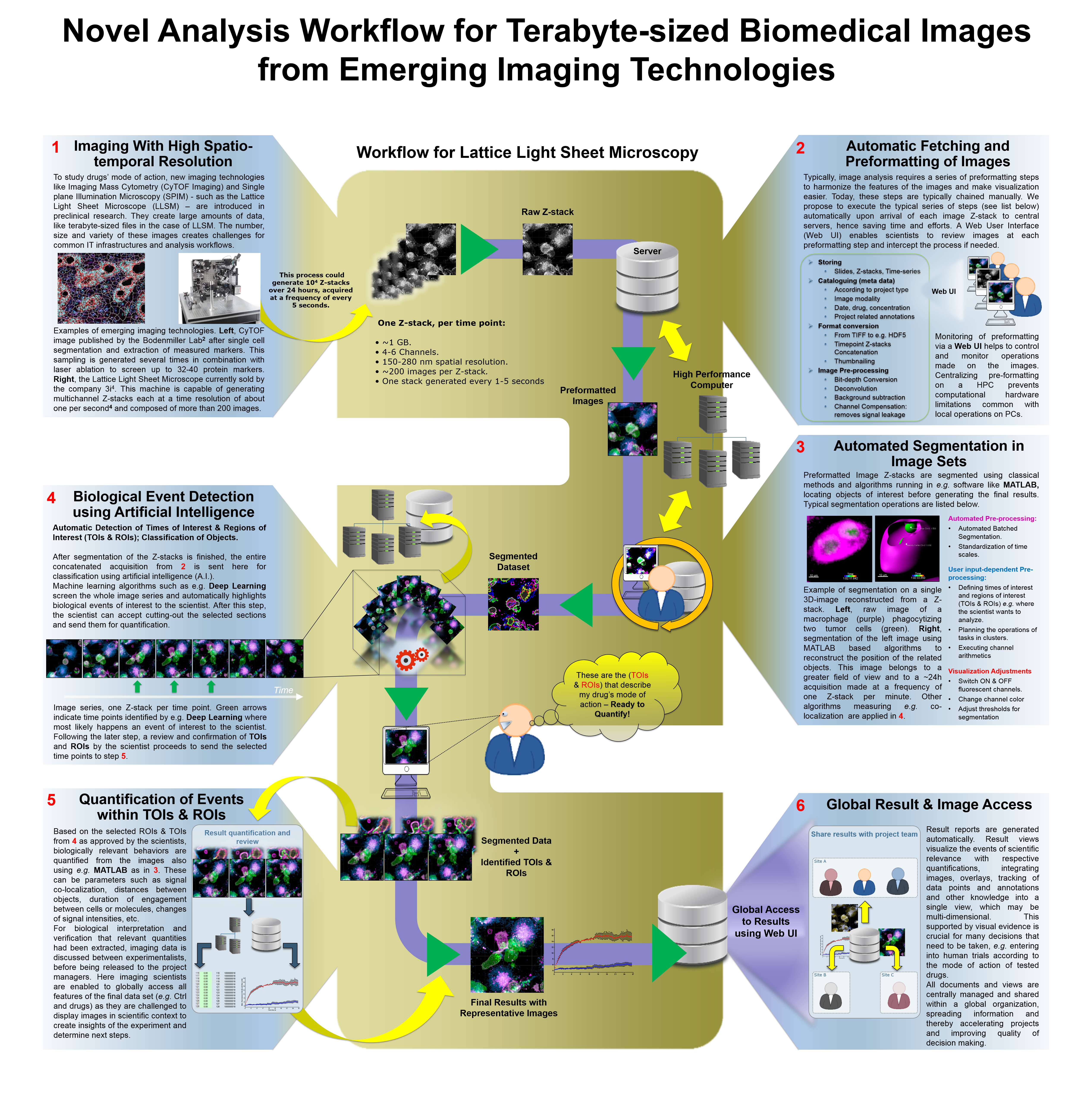
The workflow above was presented at SBI2 High Content 2016 - 3rd Annual Conference, Boston, MA, USA. You can download it at the reference section of this post (5).
In Summary:
I believe that LLSM Imaging will become a must-have technique in preclinical drug development. It will help scientists thoroughly characterize molecular mechanisms, disease models, and conditions where drugs are most efficacious.
For the same reasons, I think that pharma R&D will likely invest in LLSM imaging to address unknown mode of action mechanisms during drug development. However, the return of investment could be considerably limited without a flexible hardware infrastructure and, more importantly, a software capable of managing the huge volume of images and results created.
Such a software solution could empower scientists to focus on their experimental design instead of on the IT-related issues that preclinical image analysis currently generates.
This post was modified from my LinkedIn profile: https://www.linkedin.com/pulse/imaging-future-cancer-immunotherapies-dr-daniel-gutierrez
Further Reading
Actin Depletion Initiates Events Leading to Granule Secretion at the Immunological Synapse. Immunity 19 May 2015: Vol. 42, Issue 5, p864–876.
Highly multiplexed imaging of tumor tissues with subcellular resolution by mass cytometry. Nature Methods 02 Mar 2014: 11, 417–422.
Conditional density-based analysis of T cell signaling in single-cell data. Science 28 Nov 2014: Vol. 346, Issue 6213.
https://www.genedata.com/resources/posters/poster/?tx_infores_detail%5bresource%5d=288&no_cache=1
Congratulations @daniel-gutierrez! You received a personal award!
You can view your badges on your Steem Board and compare to others on the Steem Ranking
Do not miss the last post from @steemitboard:
Vote for @Steemitboard as a witness to get one more award and increased upvotes!