Bacterial Microcompartments: Nano-Bioreactor
Hey Steemians,
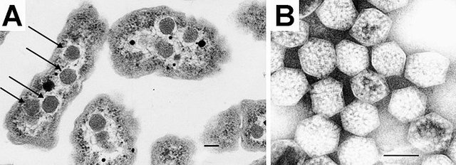
In today’s article I will be talking about bacterial microcompartments (BMC) are analogues of eukaryotic organelles; it is structure which encloses protein and enzymes inside a protein wall. The first microcompartment was discovered in 1950 using electron microscopy in cytoplasm of cyanobacteria. The structures were found to be very small in range of 100-200 nm diameter, earlier it was thought that it is viruses but later on studies with Thiobacillus it was reported to be protein shell filled with RuBisCO (ribulose-1,5-bisphophate) and it carry outs the function of carbon dioxide fixation through Calvin cycle (1). The protein wall is very selective in nature it only allows entry of certain substrates. The separation of enzymes from the cytosol environment prevents the enzymes from toxic metabolic intermediated and reduces the risk of interruption by unwanted reactions (2).
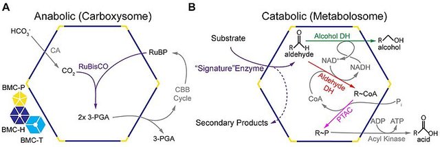
The bacterial microcompartments are of two types based on the function they carry out anabolic and catabolic. The only BMC known to carry out anabolic process is the carboxysomes; which is present in cyanobacteria. Carboxysomes BMC contains RuBisCO (fixes carbon dioxide to create two molecules of phosphoglycerate) and carbonic anhydrase (catalyzes conversion of bicarbonate to carbon dioxide) enzymes which carry out carbon dioxide fixation using Calvin cycle (3). The catabolic BMC also known as metabolosomes are expressed only when the substrate is present. The BMC are named on the basis of substrate metabolized for instance propanediol utilization (PDU-BMC), ethanolamine utilization (EUT-BMC) and fucose and rhamnose (4). The catabolic BMC differs on the basis of substrate which are metabolized but there enzymatic core are similar it consists of signature enzyme (specific to initial substrate), aldehyde dehydrogenase, an alcohol dehydrogenase and phosphotransacylase (5).
Earlier in my post I have mentioned that the BMCs are selectively permeable to certain compounds. For example in case of carboxysomes the BMC wall is permeable to RuBP, bicarbonate and phosphoglycerate on the other hand the entry of oxygen and carbon dioxide is limited (6). Another example is of propanediol utilization BMC, it is permeable to propanediol, propanol, and also to vitamin B12, while the propionaldehyde is sequestered to prevent cell damage (7). It has been proposed that the selective nature of the BMC shell proteins is due to the charges on the walls of shell proteins. The pores of carboxysomes shell protein have overall positive charges which results in entry of negatively charged bicarbonate ions (8).
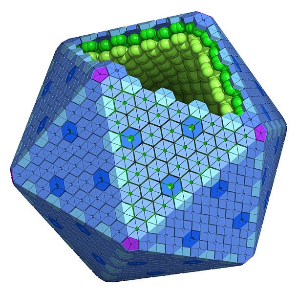
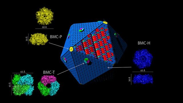
Let’s talk about the structure BMC shell and their assembly in carboxysomes and metabolosomes. The main building blocks of shell protein are BMC-H, BMC-T and BMC-P.The BMC-H is the most type of shell protein that assembles to form a cyclic homohexamer with a convex and a concave side and contains a single Pfam00936 domain (9). Next is BMC-T which is forms a trimer by the fusion of two Pfam00936 domains and it also resembles like pseudohexamer. When BMC-H and BMC-T align a pore is formed at the central symmetry axis which acts as a channel for the metabolites to enter and leave the shell (10). BMC-H and BMC-T forms the facets of shell a pentameric protein is required to cap vertices of shell. Now here BMC-P comes in the picture and forms the vertices the shell and it contains a single Pfam03319 domain which assembles to form a pentameric structure (11).
With the sequencing of the BMC shell protein, scientists started to work to solve the puzzle of assembly of BMC in carboxysomes and metabolosomes. Carboxysomes is divided into two type α-carboxysomes and β-carboxysomes. Both of them differs in the way they assembles, in β-carboxysomes the core is assembles first which is followed by the assembly of shell but in case of α-carboxysomes the assemble of core and shell takes pace together (12). The phylogenetic analysis of BMC-P protein has shown its similarity to β-carboxysomes although the assembly pathways of metabolosomes are yet to be explained experimentally (2).

So now talk about the biotechnological applications of these bacterial microcompartments. These BMCs can serve nanobioreactors. The main focus of metabolic engineering is to design pathways which for high yield of chemicals and other pharmaceuticals products. Many factors like alternative reaction pathways, accumulation of toxic intermediates and inhibitory products always results in lower yield. However in nature these conditions can be bypassed by compartmentalization, this can be seen in peroxisomes (13). Scientists have started using scaffolds made up of proteins or DNA to colocalize the enzymes in a compartment to increase product yield for example in production of glucaric acid, and mevalonate (14). Another use of these BMCs is they can be used for the delivery of metal nanoparticles, cytotoxic chemicals or cytotoxic proteins. This property of BMC has an application in cancer therapy and tumor imaging. The control release of cytotoxic drugs to targeted tumor cells and limit the risk of side effects to normal cells (15).
Summary
Bacterial microcompartments are wide spread and diverse in nature and function, made up of protein shell and can enclose protein and enzymes in it. The protein coat of these biocompartments act like a membrane as they funcion in the permeability of BMC shell. A lot of studies is being carried out to understand the nature of these cell protein and to manipulate it using mutagenesis to change the porosity and to understand the charge distribution on these cell protein. Synthetic biologists can use this microcompartments to form empty shells to carry drugs to the target site.
Carboxysomes in Motion
Reference
Kerfeld et. al., 2018 Bacterial microcompartments. (79)
Turmo et. al., 2017 Carboxysomes: metabolic modules for CO2 fixation. 234(18).
Tanaka et. al., 2008 Atomic-level models of the bacterial carboxysome shell. 319(5866),1083-1086.
Chen et. al., 2012 Designing biological compartmentalization. 22(12),662-670.

Hi @Vinamra
We have selected your post as post of the day for our DaVinci Times. Our goal is to help the scientific community of Steemit, and even if our vote is still small we hope to grow in quickly! You will soon receive our sincere upvote! If you are interested in science follow us sto learn more about our project.
Immagine CC0 Creative Commons, si ringrazia @mrazura per il logo ITASTEM.
CLICK HERE AND VOTE FOR DAVINCI.WITNESS
Keep in mind that for organizational reasons it’s necessary to use the “steemstem” and “davinci-times” tags to be voted again.
Greetings from @davinci.witness and the itaSTEM team.
Wow, I thought I knew enough about organelles. Thank you for this post, very informative!
Thanks @thethinkingdr for the appreciation. I'm glad you find it informative.
Microbes have so much for us to study and we are still to find so many new species of these with new surprises out there in the environment.
Cheers
This was new to me. When I saw the image first I thought is it like some ancient Virus trapped inside bacteria (like ancient bacteria trapped inside eukaryotes which became mitochondria.) Then I came to reality leaving my imagination :)
Thanks for your comment @dexterdev
Yeah, the microcompartments look somewhat similar to the virus head...... :)
These are just the bodies lying inside the bacterial cell and have the function in conversion pathways.
Very interesting insight. I would like to see more applications of it. It was a little hard to read but It might just be me (or more coffee is needed).
I appreciate microbiology and I would like to understand more than just the surface :P
Thanks for the comment @alexdory
I'm glad you find it interesting.